Foot and ankle pain can be frustrating, especially when it starts to impact your daily life. Whether it’s walking, standing, or just trying to get through your day without discomfort, foot and ankle issues can make even the simplest activities difficult. When pain persists and traditional diagnostic methods like X-rays don’t provide answers, an MRI (Magnetic Resonance Imaging) might be the key to uncovering what’s going on beneath the surface.
MRI is a powerful tool that can reveal hidden causes of pain by providing detailed images of soft tissues, tendons, ligaments, and bones. Let’s dive into how an MRI helps in diagnosing complex foot and ankle problems and when it might be the right option for you.
Why Diagnosing Foot and Ankle Pain Is Challenging
The foot and ankle are complex structures, made up of 26 bones, 33 joints, and more than 100 tendons, ligaments, and muscles. With so many moving parts, it’s no wonder that diagnosing the source of pain can be tricky. Issues like tendon tears, ligament damage, or small stress fractures often don’t show up on basic imaging tools like X-rays.
X-rays are great for showing bones and can help detect fractures or bone alignment issues. However, they fall short when it comes to diagnosing problems in the soft tissues—like the tendons, ligaments, and cartilage—that are often the cause of persistent pain. This is where MRI comes in as an invaluable diagnostic tool. MRI can look beyond the bones and give a clear, detailed image of what’s happening with the soft tissues inside your foot and ankle.
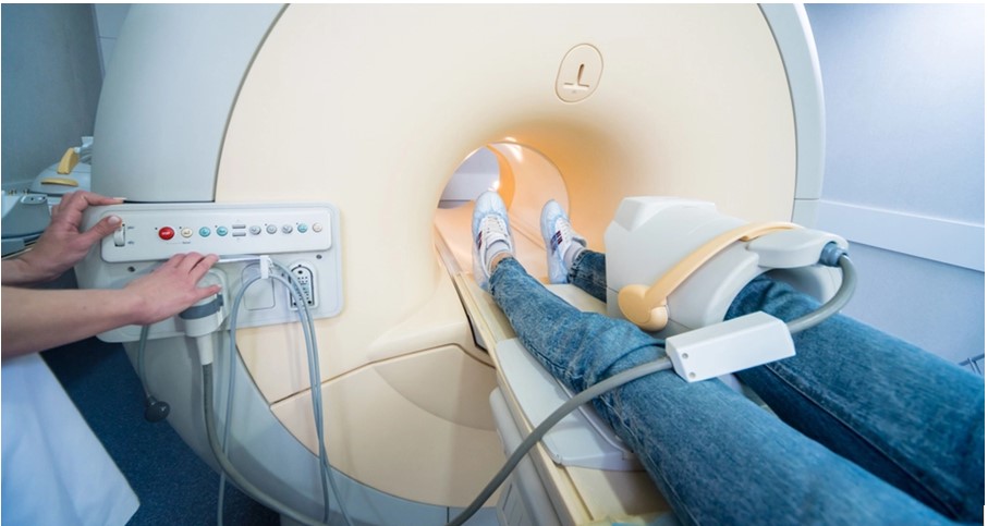
How MRI Works and Why It’s Effective for Foot and Ankle Pain
MRI uses magnetic fields and radio waves to create detailed images of the body’s internal structures. What makes MRI so effective for diagnosing foot and ankle pain is its ability to capture clear images of soft tissues—something X-rays simply can’t do. MRI can show muscles, tendons, ligaments, and cartilage in high detail, allowing doctors to see things like small tears, inflammation, or degeneration that could be causing your pain.
Another advantage of MRI is that it’s completely non-invasive and doesn’t use radiation, making it a safe option, even for long-term or repeat use. You just lie still while the machine takes images, and the results can provide crucial insights into the cause of your discomfort.
Common Foot and Ankle Issues Diagnosed with MRI
Here are some of the most common foot and ankle problems that can be accurately diagnosed with an MRI:
Tendon Injuries (Achilles Tendon, Posterior Tibial Tendon)
Tendon injuries are a common source of ankle pain. Whether it’s tendonitis or a tear, MRI can clearly show the extent of inflammation or damage. For example, Achilles tendon injuries are frequent in active individuals, and MRI helps identify whether the injury is a minor strain or something more severe, like a partial or full tear.
Ligament Damage (Ankle Sprains)
Ankle sprains often involve torn or overstretched ligaments. While some sprains heal on their own, severe cases may require medical intervention. MRI can reveal the full extent of ligament damage, ensuring that you get the proper treatment to avoid long-term instability or chronic pain.
Stress Fractures
Stress fractures are tiny cracks in the bone that can develop from overuse, such as in athletes or people who are on their feet a lot. These small fractures are often missed on X-rays, but MRI can detect them, allowing for early treatment and preventing further complications.
Plantar Fasciitis
Plantar fasciitis is a common cause of heel pain, resulting from inflammation or degeneration of the plantar fascia, the thick band of tissue that runs along the bottom of your foot. MRI can provide a clear picture of the affected area, helping doctors to tailor the right treatment plan.
Cartilage and Joint Issues (Osteoarthritis, Gout)
In conditions like osteoarthritis or gout, MRI can help doctors assess the condition of the cartilage in your joints and detect fluid buildup, which is often associated with these types of arthritis. This information is crucial in managing pain and slowing the progression of joint damage.
Neuroma or Nerve Compression (Morton’s Neuroma)
Morton’s neuroma occurs when tissue thickens around a nerve in the foot, usually between the toes, causing sharp pain. MRI can confirm the diagnosis by clearly showing the compressed or inflamed nerve.
Benefits of Using MRI for Foot and Ankle Pain Diagnosis
The benefits of using MRI for diagnosing foot and ankle pain are numerous. First and foremost, MRI can catch soft tissue injuries early, allowing for prompt treatment before the problem worsens. Early detection of injuries like tendon tears or ligament strains can make a huge difference in your recovery time and overall outcome.
MRI also provides an accurate diagnosis, which leads to better treatment plans. With a clear understanding of what’s causing your pain, your doctor can develop a targeted approach to treatment, whether that involves physical therapy, medication, or surgery.
Another key advantage of MRI is that it’s non-invasive and pain-free. You won’t have to undergo any uncomfortable procedures to get a clear picture of what’s going on inside your foot or ankle.
Finally, MRI is not only useful for diagnosis but also for monitoring recovery. After an injury, MRI can track the healing process, helping your doctor adjust your treatment plan as needed and ensuring that you’re on the right path to full recovery.
When Should You Consider an MRI for Foot and Ankle Pain?
You might wonder when it’s time to consider an MRI. Generally, an MRI is recommended if you’ve been dealing with chronic pain that doesn’t improve with rest, ice, or other conservative treatments. If you’ve had an injury and your pain persists despite initial treatment, an MRI might be necessary to determine the full extent of the damage.
If soft tissue damage is suspected but can’t be confirmed with an X-ray or physical exam, an MRI is the next step. For post-injury assessments, such as after a bad sprain, MRI can help ensure that everything is healing properly and that no additional issues are lurking beneath the surface.
If your X-rays come back normal but you’re still in pain, an MRI might reveal hidden issues like stress fractures or nerve compression that aren’t visible on standard imaging.
How MRI Results Influence Treatment Decisions
Once the MRI results are in, your doctor will be able to make informed decisions about the next steps in your treatment. If the damage is minor, conservative treatments like physical therapy or rest might be enough to resolve the issue. In more severe cases, such as tendon tears or stress fractures, your doctor might recommend surgical intervention.
MRI results also help prevent re-injury by guiding your recovery process. Whether that involves recommending specific exercises, advising on proper footwear, or adjusting your activity level, MRI provides the detailed information needed to keep you on the right track.
Conclusion
MRI plays a crucial role in diagnosing foot and ankle pain, especially when other imaging methods fall short. By providing detailed images of the soft tissues, tendons, ligaments, and bones, MRI helps doctors identify the root cause of your discomfort, allowing for more precise treatment plans. If you’re dealing with unresolved foot or ankle pain, it might be time to consider an MRI.
At Upright MRI of Deerfield, we specialize in providing accurate, comfortable MRI scans to help diagnose the source of your pain. Reach out to us today to schedule your appointment and take the first step toward finding relief and getting back on your feet.